


Case Report
Victor Hugo Ramirez Lopez and Hector Mauricio Serrano Reyes
Correspondence Address :
Victor Hugo Ramirez Lopez
Carlos Chagas State Hospital
General
Surgery Service
Rio de Janeiro, RJ
Brazil
Received on: November 20, 2020, Accepted on: May 12, 2023, Published on: May 15, 2023
Citation: Victor Hugo Ramirez Lopez, Hector Mauricio Serrano Reyes (2023). Terminal Ileum Perforation By Burkitt Non-Hodgkin Lymphoma In A Pediatric Patient: A Case Report
Copyright: 2023 Victor Hugo Tamirez Lopez, et al. This is an open-access article distributed under the terms of the Creative Commons Attribution License, which permits unrestricted use, distribution, and reproduction in any medium, provided the original author and source are credited.
Lymphoma is the third most frequently diagnosed neoplasm in children and can be primary extra nodal, if there is no or minimal nodal lymph node involvement; or secondary involvement, when the disease is already advanced, this being significantly more common. The peak age for Burkitt lymphoma in children is 5-15 years, with more cases in boys than in girls. The clinical manifestation is related to the location of the primary tumour. The most common presentation of primary abdominal Burkitt lymphoma is reported to be abdominal pain and can present itself urgently with an acute exacerbation caused by intussusception, appendicitis or perforation like our case report of a an 9-year-old male child who sought the Emergency Care Unit (UPA), with a report of an intestinal obstruction evolving to perforation and peritonitis, Emergency surgical procedure was developed, which served both as a therapy and to investigate the disease, as it allowed the diagnosis of Non-Hodginlymphoma. Primary gastrointestinal tumours in children are rare. Non- Hodgin lymphoma remains the most common malignancy of the gastrointestinal tract in children but with the proper diagnosis and treatment the patients would have a high rate of good clinical response.
Keywords: Perforated Ileum, Lymphoma, Burkitt lymphoma, Non-Hodgin lymphoma, Intestinal obstruction
Introduction
Intestinal perforation can be caused by several reasons, such as (i) the ingestion of elongated and pointed foreign bodies [1], which causes trauma directly to the intestinal wall; (ii) diseases such as appendicitis, diverticulitis, lymphoma etc.; (iii) biliary calculus or infection; (iv) radiation injuries; and (v) iatrogenic causes, for example, from an endoscopy (but this is rare). In about 83% of cases, the ileal loops are impaired [2]. Lymphoma is the third most frequently diagnosed neoplasm in children and adolescents up to 15 years of age. In this demographic, about 60% of cases involve non-Hodgkin’s lymphoma (NHL) and the rest are due to Hodgkin’s lymphoma (HL). Burkitt-type NHL is prevalent in Brazil, with abdominal presentation being the most usual [8]. As stated in the medical literature, extranodal involvement affects about 40% of patients with lymphoma [3]. Lymphomas can be primary extranodal, if there is no or minimal nodal lymph node involvement [4,5]; or secondary involvement, when the disease is already advanced, this being significantly more common [3,6]. It is most commonly found in Non-Hodgkin’s Lymphoma (NHL), present in 25% of cases [7]. The late diagnosis of lymphoma can take major proportions, such as intestinal perforation, as occurred in the analyzed patient. In general, patients with intestinal perforation seek emergency services with acute symptoms in the abdomen, such as tachycardia, abscess, fistula, abdominal pain on superficial or acute palpation, nausea, vomiting, fever, peritonitis, intestinal obstruction, and gastrointestinal bleeding.
Pain from intestinal perforations can extend to the hip and groin; and the leakage of intestinal contents is capable of causing peritonitis, abscess formation in the abdomen and even a severe septicemia. If not properly treated, this condition can cause serious problems to the patient, leading to death.
Studies have been dedicated to examining the behavior of NHL in children and youth. However, these are still scarce. Based on these works, it is noted that the incidence of NHL increases uniformly as the child ages, being a disease that rarely affects children under two years of age [8,13,14]. In the present study, the patient is nine years old, thus fitting the expected standard, since NHL can also affect young individuals up to 15 years old. It is possible that the occurrence of an infectious etiology in the pathogenesis of Burkitt’s lymphomas, mainly related to the presence of the Epstein-Barr virus (VEB) [9], is able to explain, in part, the appearance of this disease in younger individuals, mainly if they come from more disadvantaged economic classes, since, as a rule, this population is exposed earlier to infectious agents [8]. Lymphomas in children and youth, especially those classified as Burkitt’s, have a tendency to develop at an accelerated rate, and may already be very advanced at the time of diagnosis, which requires immediate treatment, in order to avoid serious damage to the sick. These lymphomas can be treated with chemotherapy, since it has the potential to destroy a large percentage of lymphoma cells in a short period of time. In this scenario, the concern relates to the fact that lymphomas can cause tumor lysis syndrome, which is a side effect that can occur when the contents of dead cells enter the bloodstream, which can cause problems in other organs. As a way to avoid this consequence, doctors usually prescribe, in addition to the drugs, the intake of a large amount of fluids during treatment.
NHLs are neoplasms, which can vary significantly, both in terms of growth rate and response to treatment. Furthermore, it is believed that children diagnosed with lymphoma in early stages I or II, have the disease more widespread than what is presented by the tests performed, whether laboratory or imaging. As a result, only the application of specific treatments, such as surgery or radiation therapy, is considered effective for curing the disease. Thus, chemotherapy is considered a useful tool for the treatment of lymphomas in patients of all ages, including children, varying according to stage and type of disease. When patients are treated up to stage II, as in the patient studied here, the survival rate reaches up to 95% in 5 years [10-12].
The treatment and efficient prognosis of a person with NHL depends on the type and stage of the disease, which can be verified, initially, with imaging exams. Therefore, the use of positron emission tomography or computed tomography has been shown to be the most effective means of determining the stage and type of lymphoma.
Once the presence of lymphoma is identified in the patient, it is necessary to carry out tests to verify the extent of the disease in the body. Moreover, it is advised that the diagnosis of NHL be carried out based on ganglion biopsy. This is extremely important, since there are several subtypes of NHL, some of which are slow- growing and have discrete symptoms, while others may present rapid growth and exacerbated symptoms.
Case Report
G.S.S, male, 9 years old, from the Emergency Care Unit (UPA), presented abdominal pain, mainly in the right iliac fossa, associated with nausea, fever and vomiting, lasting approximately 24 hours. Furthermore, he was tachycardiac and with slight tachypnea. Physical examination returned signs of peritoneal irritation.
Laboratory tests showed significant leukocytosis (33,200) with neutrophilia. Tomography of the total abdomen presented pneumoperitoneum and presence of a moderate amount of free intracavitary fluid.
The results indicated urgent exploratory laparotomy. After performing the necessary procedures, according to the surgical report, the patient had a large blockage in the pelvis, with fibrin and an area of necrosis compromising the terminal ileum with its perforation, which was undone (Figures 1 and 2). With that, an ileocolectomy with end-to-end anastomosis was performed and the specimen was sent to histopathology (Figures 3 and 4).
The intraoperative finding revealed the presence of pelvic peritoneal block with the identification of perforation of the terminal ileum, but with cecal appendix without changes. From that, ileocolectomy was performed and the specimen sent for biopsy. Histopathological analysis of the surgical specimen showed the presence of Burkitt-type B-cell non-Hodgkin’s lymphoma (Figure 5).
In the postoperative period, the patient stayed at the pediatric Intensive Care Center (ICU), where he remained for five days for the purposes of clinical stabilization. On the third postoperative day, a trial liquid diet was started, accompanied by intravenous hydration and antibiotic therapy, which provided a satisfactory evolution. On the fifteenth day, the patient was discharged and was referred to the general surgery outpatient clinic.
The result of the anatomopathological examination revealed the existence of intestinal lymphoma with extensive necrosis and fibrosis in the perforation area. The diagnosis was complemented with the immunohistochemical analysis of Burkitt-type B-cell non-Hodgkin’s lymphoma.
Discussion
In the present study, we analyze a 9-year-old male child who presented a disease in a localized way, that is, stage II. The patient had several complaints that indicated the presence of intestinal perforation, such as acute abdominal pain, nausea, fever and vomiting episodes.
The peak age for Burkitt lymphoma in children is 5-15 years, with more cases in boys than in girls (3.9:1.1) [17]. BL is characterized by dysregulation of the c-MYC oncogene [17], Deregulation of c-MYC prevents B-cell differentiation and has oncogenic activity [18]. Cytogenetic evidence of c-MYC rearrangement is the gold standard for the diagnosis of BL [19]. Three major clinical types of Burkitt lymphoma exist: (1) the endemic form, which is a common childhood malignancy strongly associated with the Epstein-Barr virus (EBV), (2) the non-endemic
(sporadic) form which is rare, and (3) immunodeficiency-related BL, mostly seen in AIDS patients. The sporadic form typically presents itself as extranodal disease. The clinical manifestation is related to the location of the primary tumour. According to various studies, the most common presentation of primary abdominal Burkitt lymphoma is reported to be abdominal pain (85%), followed by abdominal swelling, vomiting, constipation, diarrhoea, melena, and rectal bleeding [20]. Symptoms can present themselves urgently with an acute exacerbation caused by intussusception, appendicitis, perforation, or bowel obstruction [18]. About 14% of Burkitt lymphoma tumours can arise in the bone marrow. This variety is called Burkitt cell leukemia [21]. CNS involvement is diagnosed in 8.8% of patients with Burkitt lymphoma/leukemia [17].
It is important to note that non-surgical procedures are not shown to be very effective methods for diagnosing abdominal NHL in children and youth, as they have a low morbidity rate [15]. As a result, urgent surgery was indicated. Considerable necrotic area and ileum perforation were found, and an attempt was made to repair the damage immediately with the surgical procedure. The removed surgical specimen was sent for histopathological analysis, which verified the presence of Burkitt-type B-cell NHL. Furthermore, pediatric NHL, as a rule, is an aggressive tumor with a high potential for cell proliferation. In this sense, it is important to use a technique capable of collecting the ideal quantity and quality of pathological material, as a way of ensuring that the tests are properly and timely performed, thus enabling a timely diagnosis, which is not always possible by means of non- surgical procedures.
Primary gastrointestinal tumours in chil- dren are rare. Non-Hodgin lymphoma remains the most com- mon malignancy of the gastrointestinal tract in children. In most cases the histologic subtype of the setumoursis Burkitt- lymphoma. It can present with an acute peritonitis caused by Intestinal perforation. Burkittlymphomais a highly aggressive, rapidly growing neoplasm. However, if treatment is started in time and there is no unfavorable riskfactors, the good clinical res- ponse with 3 years event free survival (EFS) is more than 95% Combination chemotherapy. According to standardized proto- cols is the best current standard of care. How was presented in the reported case the difference is in the diagnostic suspicion and prompt treatment for the best posible prognosis.
1. Ziter FM Jr. Intestinal perforation in adults due to ingested opaque foreign bodies. Am J Gastroenterol. 1976;66:382-385.
2. Singh RP, Gardner JA. Perforation of the sigmoid colon by swallowed chicken bone: case reports and review of literature. Int Surg. 1981;66:181-183.
3. Lee WK, Lau EWF, Duddalwar VA, Anthony J Stanley, Yvonne Y Ho. Abdominal manifestations of extranodal lymphoma: spectrum of imaging findings. AJR Am J Roentgenol. 2008;191 (1):198-206.
4. Leite NP, Kased N, Hanna RF, et al. Cross-sectional imaging of extranodal involvement in abdominopelvic lymphoproliferative malignancies.
Radiographics. 2007;27 (6):1613-1634.
5. Silva Neto MM, Jalil EM, Araujo IBO. Extranodal non-Hodgkins lymphomas in Salvador, Brazil: clinical aspects and histopathological classification according to the WHO-2001 guidelines. Rev Bras Hematol Hemoter. 2008;30:36-40.
6. Kwee TC, Nievelstein RAJ, Torigian DA. Role of structural imaging in lymphoma. PET Clin. 2012;7(1):1-19.
7. Fajardo L, Ramin GA, Penachim TJ, Martins DL, Cardia PP, Prando A. Manifestações abdominais do linfoma extranodal: ensaio iconográfico. Radiologia Brasileira. 2016;49(6):397-402.
8. Márcia Ferreira Pedrosa MF; Pedrosa, F; Lins, MM; Net, NTP; Gilliatt, HF. Linfoma não-Hodgkin na infância: características clínico-epidemiológicas e avaliação de sobrevida em um único centro no Nordeste do Brasil. J. Pediatr. 2007;83(6).
9. Gutiérrez MI, Bhatia K, Barriga F, et al. Molecular epidemiology of Burkitt’s lymphoma from South America: differences in breakpoint location and Epstein-Barr virus association from tumors in other world regions. Blood. 1992;79(12):3261-3266.
10. Torricelli, FCM, Lopes, RI, Dias, AR; et al. Linfoma ileal primário como uma causa de intussuscepção ileocecal recorrente. Rev bras. colo-proctol. 2008;28(2).
11. Stovroff MC, Coran AG, Hutchinson RJ. The role of surgery in American Burkitt’s lymphoma in children. J Pediatr Surg. 1991;26(10):1235-1238.
12. Armitaje JO. Treatment of non-Hodgkin’s lymphoma. N Engl J Med. 1993;328(14):1023-1030.
13. Sandlund JT, Fonseca T, Leimig T, et al. Predominance and characteristics of Burkitt lymphoma among children with non-Hodgkin lymphoma in northeastern Brazil. Leukemia. 1997;11:743-756.
14. Bittencourt AL, Mendonça N, Cordeiro CO, et al. Linfoma maligno não- Hodgkin na infância: estudo clínico-patológico de 70 casos. J Pediatr (Rio J). 1987;62(6):259-262, 265-266.
15. Aguiar, AA; Lima, LC; Araújo, CC; Rodrigo Melo Gallindo, RM. Pediatric abdominal non-Hodgkin’s lymphoma: diagnosis through surgical and non- surgical procedures. J. Pediatr (Rio J). 2019;95(1):54-60.
16. Zelenetz AD, Gordon LI, Wierda WG, Abramson JS, Advani RH, Andreadis CB, et al. Non-Hodgkin’s lymphomas. Version 2.2015. In: National Comprehensive Cancer Network (NCCN), clinical practice guidelines in oncology (NCCN guidelines). Washington: NCCN; 2015.
17. PDQ® PediatricTreatment Editorial Board. PDQ childhood non-Hodgkin lymphoma treatment. Bethesda, MD: NationalCancerInstitute; Updated 03/30/2018.
18. Gualco G Weiss LM Harrington WJ Bacchi CE Jr. Nodal diffuselarge B-celllymphomas in children and adolescents: immunohistochemical
expression patterns and C-Myc translocation in relation to clinical outcome. Am J SurgPathol. 2009;33(12):1815-1822.
19. Allen CE, Kelly KM, Bollard CM. Pediatric lymphomas and histiocytic disorders of childhood. Pediatric Clinics. 2015;62(1):139-165.
20. Ghoroubi J, Mirshemirani A, Kouranloo J, Nazari S. Abdominal Burkitt’s lymphoma in children. Iranian Journal of Pediatric Surgery. 2015;1(1):28-33.
21. Mbulaiteye SM, Biggar RJ, Bhatia K, Linet MS, Devesa SS. Spora- dic childhood Burkitt lymphoma incidence in the United States du-
ring 1992 2005. Pediatr Blood Cancer. 2009;53(3):366-370.
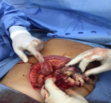
Figure 1 - Perforated terminal ileum

Figure 2 - Ileal necrosis

Figure 3 – Surgical specimen

Figure 4 - Surgical specimen

Figure 5 - Burkitt Lymphoma

Clinical Microbiology & Case Reports

Women's Health & Gynecology

Journal of Global Diabetes & Clinical Metabolism

Journal of Alzheimers Parkinsonism and Dementia

Journal of Dental and Oral Health

Journal of Ophthalmology & Visual Neuroscience

Cancer Science: Open Access

Journal of Immunology, Infection & Inflammatory Diseases

Journal of Prevention & Treatment of HIV/AIDS

Journal of Primary Health Care & General Practice

Journal of Blood Disorders Symptoms and Treatments

Journal of General and Emergency Medicine

Journal of Clinical Anesthesia and Pain Medicine

Journal of Applied Physics & Nanotechnology

Journal of Advances in Breast Cancer Research and Development

Interdisciplinary Journal of Gastroenterology, Hepatology and Endoscopy
Journal of General Surgery

Journal of Global Vaccines R&D

Journal of Integrative Pediatric Healthcare: Open Access

SOA-Clinical & Medical Case Reports, Reviews

Interdisciplinary Journal of Bone Marrow & Stemcell

Interdisciplinary Journal of Nursing and Critical Care

Global Journal of Internal Medicine

Journal of Integrative Food Sciences & Nutrition

Integrative Clinical Cardiology

Clinical Dermatology and Investigations

Molecular Medicine: Current Aspects

Open Access Journal of Public Health (ISI Indexed)

Annals of Clinical Research and Trials

Global Chemical Engineering & Process Technology

Integrative Plant Science & Molecular Biology

Interdisciplinary Journal of Translational Science

Integrative Neurology and Stroke

Journal of Vascular Diseases and Therapy

Integrative Trauma and Emergency Medicine

SOA-Otolaryngology-ENT Research

Global Urology and Nephrology

Journal of Pregnancy and Newborncare

SOA-Applied Biotechnology & Bioengineering

Journal of Obesity Treatment and Weight Management

SOA Medical Research & Clinical Practice

Integrative Clinical Pathology

Global Toxicology

Dairy Science, Veterinary and Animal Husbandry

SOA Radiology & Radiation Therapy

Global Analytical Techniques & Pharmaceutical Research

Abnormal Psychology and Clinical Psychiatry

SOA Archives of Pharmacy & Pharmacology

Global Nutrition and Dietetics

Global Hydrologic Engineering Research

Global Physical Medicine & Rehabilitation Practices

Integrative Oral care and Craniofacial Research
Integrative Ecology and Environmental Sciences

Open Access Journal of Sociology
Interdisciplinary Journal of Fisheries and Aquaculture research
Biomedical Research and Clinical Studies

Clinical Epidemiology and Healthcare

Molecules Research & Analysis
Journal of Polymers Research


 Open Access
by Scient Online is licensed.
Open Access
by Scient Online is licensed.

